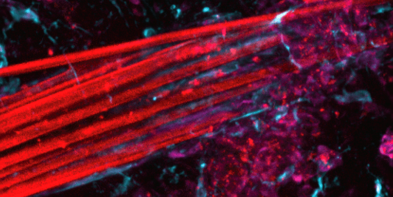
A team at ETH Zurich is pushing the boundaries of what can be achieved with neurostimulation and neuronal recording technology thanks to the super-flexible electrode bundles they have developed. Rather than traditional electrodes, which can cause some damage to brain tissue when they are inserted, the fibers can seamlessly integrate into the web of dendritic filaments with no harm done.
Electrodes are a vital part of brain-computer interfaces (BCIs) and deep-brain stimulation (DBS) devices. In DBS, a pacemaker-like component instructs the electrode to deliver bursts to electricity to a region of the brain to help relieve the symptoms of conditions like Parkinson’s disease and obsessive-compulsive disorder. In BCIs, electrode arrays record brain activity that can then be translated by a computer system into, for instance, speech or movement. Perhaps the most well-known developer of BCIs is Elon Musk’s Neuralink.
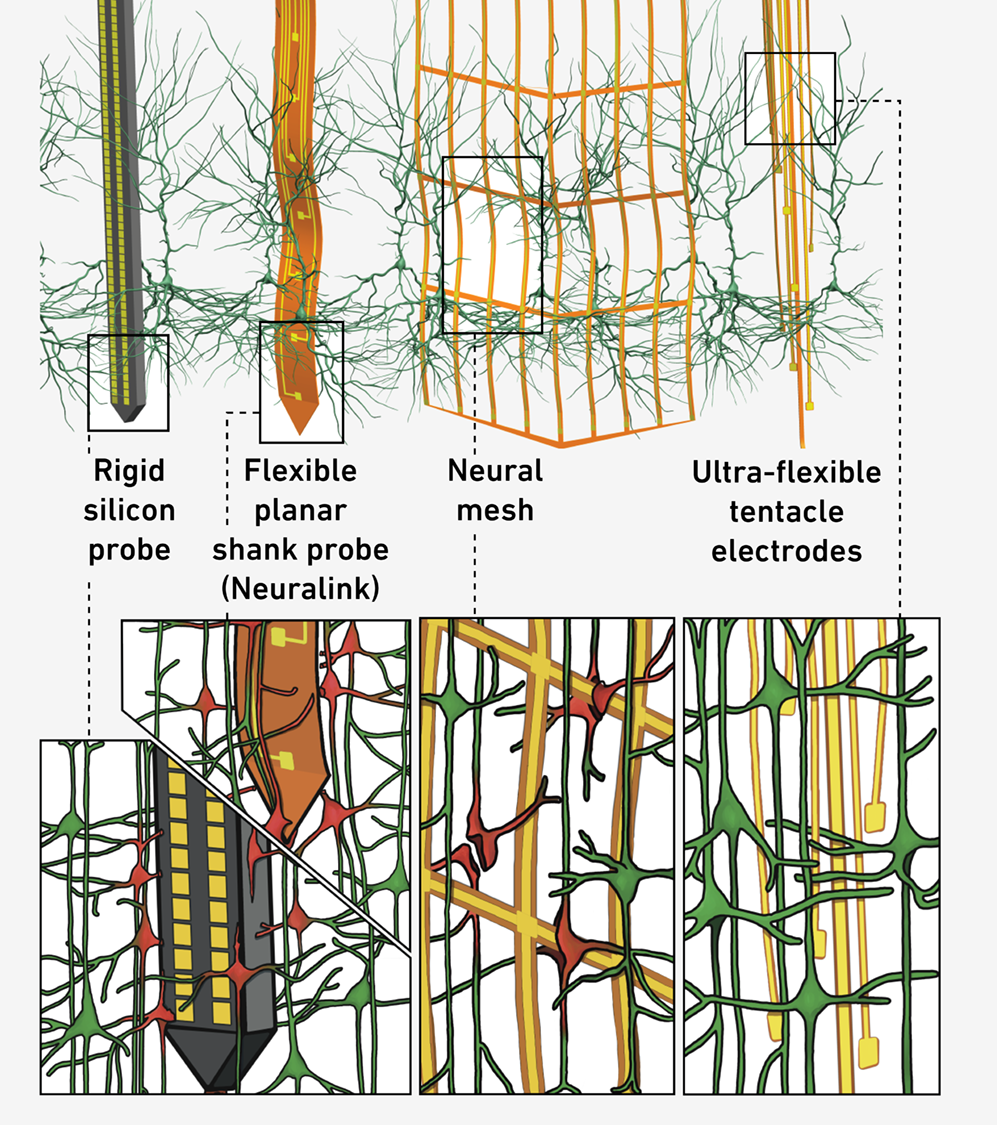
Neuralink’s implant uses a somewhat flexible probe that is an improvement on more rigid, silicon-based probes. Others are working with electrode meshes that are more biocompatible still; but when Mehmet Fatih Yanik, Professor of Neurotechnology at ETH Zurich, first wanted to experiment with brain recording tech a decade ago, it quickly became clear that a new solution was needed.
I'm always telling my students that one day they may be putting this thing into my brain.
Professor Mehmet Fatih Yanik
Yanik told IFLScience that his lab brings together neuroscientists and engineers who, over time, have “built the technology that we wanted to have". By combining their expertise, the team has now unveiled a brand-new type of electrode: a spaghetti-like bundle of extremely fine gold fibers encapsulated in a polymer. By recapitulating, as closely as possible, the structure of the tissue into which you want to insert the electrode, you can make that insertion as gentle as possible while also maximizing the potential applications of the technology.
“If you think about the neurons themselves, the cell body is about 20 microns, 10 microns,” Yanik told IFLScience. “And then you have these dendritic arbors, which are on a few-micron scale. And when you try to [insert] most of these existing technologies today, either silicon or even polymer ones, what happens is you basically are trying to go through a tissue segment, and you have these branches, like tree branches. And what you are doing is you are basically chopping it off.”
The spaghetti fibers, by contrast, can integrate into these “tree branches” without causing damage.

So far, so good. But with something so delicate and flexible, perfecting the insertion process took a lot of time: “if you wanted to get into, you know, just a few millimeters below the surface of brain, they just peel off, you know?” Lead author Tansel Baran Yasar, along with colleagues Peter Gombkoto and Wolfger von der Behrens, spent many months on this problem.
The key, it emerged, was inserting the bundles slowly. Postdoctoral researcher Angeliki D. Vavladeli has been continuing to analyze the effects of the insertion on the surrounding brain tissue, and Yanik told us that “it really looks unbelievable.”
“The tissue around is very healthy, like starting from the very next neuron there, you know, there's no layer of some junk or anything. It's very beautiful.”
For the new study, the team tested four bundles of 64 fibers each in the brains of rats. The electrodes were wired up to a recording device that was attached to the rats’ heads, so the animals could move about freely while their brain activity was logged. They demonstrated how neuronal recordings could be performed over long timescales and with good signal quality, as the probes get very close to the nerve cells.
Theoretically, many more than 64 fibers could be included in future iterations, facilitating simultaneous recordings from multiple brain areas and possibly entire brain networks.
“A big, significant effort of our research is right now focused on trying to understand how the brain is representing information […] and how the different brain areas communicate with each other,” Yanik told IFLScience.
Among the many further advances the team is working on right now, under the leadership of Eminhan Ozil they’re hoping to engineer the electrode tentacles to include “barcodes” that can tell you precisely which bundle a signal is coming from to a 60-micron resolution – that means “you can tell in the cortex which cortical layer you are [in].” There are also plans to try inserting many more bundles into the brain at once, with the aim of going up to 3,000 separate recording channels – this is currently under development in the lab, headed up by Alexei Vyssotski and Houman Javaheri.
And as well as recording neuronal activity, there’s the possibility of using these electrodes as minimally invasive neurostimulators too.
Some time soon, testing will need to move from animal models into humans. A project with collaborators at University College London is already underway, which aims to trial the electrodes as a precision diagnostic tool for people undergoing brain surgery for epilepsy.
Yanik knows that, as one of the research leads, the time may come for him to experience the innovation first-hand, and he’s prepped the team accordingly.
“I'm always telling my students that one day they may be putting this thing into my brain,” Yanik told IFLScience. “So please make sure it is really nice!”
The study is published in the journal Nature Communications.


